

This study evaluated compliance with NICE diagnostics guidance DG42 and assessed interpretation and reporting terminology for MMR immunohistochemistry in line with BAGP 2020 recommendations.

Poster 2: Endometriosis, an uncommon histological finding in the bladder
Bladder Endometriosis accounts for<2% of endometriosis in women in 2nd and 5th decade of life. Luminal endometriosis in bladder is a very rare clinical presentation, which presents as a bluish polypoid lesion seen in cystoscope. This is a mimicker of a neoplasm in bladder wall and histopathology is the gold standard in diagnosis. Endometrial glands will be seen within endometrial stroma. Immunohistochemical staining with CK7,CD10 and ER will be helpful.

Poster 3: CTNNB1-mutated ovarian tumour with biphasic morphology and conflicting immunohistochemistry: A case report
An unusual ovarian tumour with biphasic morphology and confirmed CTNNB1 mutation on molecular testing. Histology demonstrates a low-grade endometrioid component resembling borderline endometrioid adenofibroma admixed with grade 1 endometrioid adenocarcinoma, alongside a grade 3 endometrioid adenocarcinoma. The high-grade component shows solid basaloid morphology with extensive geographic necrosis and nuclear β-catenin immunoreactivity associated with the above mutation. An additional noteworthy feature is diffuse WT1 positivity in the borderline and low-grade endometrioid component, an unexpected finding and potential diagnostic pitfall in endometrioid neoplasia given its usual association with serous tumours. This case highlights the morphological and immunophenotypic heterogeneity of ovarian endometrioid neoplasms and emphasizes the diagnostic value of nuclear β-catenin expression and molecular confirmation of CTNNB1 mutation in this tumour subtype.
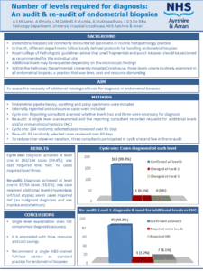
Poster 4: An audit and reaudit on endometrial biopsies- Number of levels required for diagnosis
Audit of endometrial biopsies comparing multi-level vs single-level examination. Single-level diagnosis was sufficient in >98% of cases with minimal need for additional levels of IHC. This approach maintains diagnostic accuracy while reducing time, workload and cost.

Poster 5: Acute erythroid leukemia causing fetal demise at 25 weeks gestation without associated down syndrome: A case report
This report highlights a case of congenital pure erythroid leukemia in the placenta of a 25-week stillborn fetus. Histology shows infiltration of fetal vascular spaces by erythroid blasts. Karyotype showed no evidence of trisomy 21; an uncommon occurrence in congenital leukemias.

Poster 6: Isolated giant vulvar neurofibroma in a child and the essential diagnostic role of immunohistochemistry in a resource-limited setting
This case demonstrates the essential diagnostic value of immunohistochemistry in pediatric gynecological pathology. Limited access to this tool in our setting remains a major obstacle to establishing an accurate diagnosis and providing efficient care for patients.
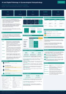
Poster 7: AI and digital pathology in gynaecological histopathology: a rapid scoping review of clinical tasks, performance and readiness
Can AI replace the pathologist’s eye in gynaecological cancer diagnosis? Our scoping review of 15 studies finds AI closest to clinical use for endometrial cancer detection — but validation gaps remain a key barrier.

Poster 8: Utility of placental examination in unravelling intrauterine infections – Foetal autopsy analysis
Placental examinations play a pivotal role in uncovering intrauterine infections, While autopsy complements by determining the exact cause of death. Together, they explain adverse outcomes, guide future clinical care and strengthen surveillance.

Poster 9: Case series of abnormal uterine bleeding presented to a secondary care hospital in Sri Lanka
Abnormal uterine bleeding is bleeding or spotting between periods, postcoital bleeding, abnormally heavy periods or irregularity in timing of periods. Any bleeding in postmenopausal age is considered abnormal. This can be due to an endometrial pathology like, polyps, hyperplasia, atypical hyperplasia ,carcinoma ,submucosal leiomyoma, endometrial stromal tumours, endometritis, exogenous hormonal effect or pregnancy related bleeding. Causes that will not appear in endometrial biopsies are adenomyosis and endometriosis. When above significant causes excluded, bleeding is due to hormonal imbalance causing dysfunctional uterine bleeding associated with non-ovulatory or ovulatory cycles. In this case series presented to secondary care hospital, most prevalent presentation is dysfunctional uterine bleeding with secretary pattern of endometrium.
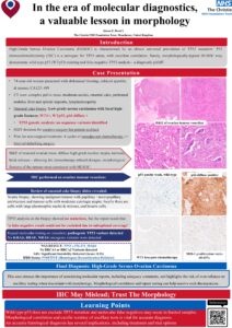
Poster 10: In the era of molecular diagnostics, morphology remains key: A valuable lesson
Morphologically high-grade serous carcinoma but wild-type p53 and negative for TP53 mutation? Trust morphology and question ancillary test results – WT p53 doesn’t rule out mutation and molecular tests can be false negative in suboptimal samples.

Poster 11: An audit on hysterectomies; histopathologic analysis of non-oncological etiologies for abnormal uterine bleeding – Experience of a tertiary care hospital, Sri Lanka
This study evaluates hysterectomies performed during 2024-2025 within a tertiary care center, Sri Lanka, for non-malignant causes, presented with abnormal uterine bleeding (AUB). This study focuses on underlying structural pathologies and the agreement between preoperative and histopathological diagnoses.

Poster 12: Müllerian adenosarcoma of the uterine corpus: report of three cases with distinct clinicopathological features
Müllerian adenosarcoma (MA) is a rare biphasic uterine neoplasm composed of of malignant mesenchymal and benign epithelial components. We report 3 cases with distinct clinicopathologic features and outcomes. A low-grade MA showed indolent behavior without recurrence. In contrast, tumors with high-grade features, including stromal overgrowth, marked atypia, high mitotic activity, myometrial invasion, and heterologous elements, were associated with more aggressive clinical courses, including early recurrence and death. These findings highlight the prognostic relevance of histologic features, particularly stromal overgrowth and high-grade components, which should be carefully assessed and reported to guide risk stratification and clinical management.
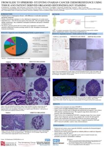
Poster 13: From slide to spheroid: Studying ovarian cancer chemoresistance using tissue and patient derived organoid histopathology staining
We successfully established patient-derived ovarian cancer organoid lines that preserve key histopathological characteristics of the originating tumours and were generated within clinically relevant timeframes compatible with treatment decision-making. Importantly, participating patients demonstrated strong engagement and willingness to support development of personalised experimental models aimed at improving future ovarian cancer care.

Poster 14: Small cell ovarian carcinoma hypercalcaemic type (SCCOHT): histopathological fidelity between tissue and organoid
We demonstrate successful generation of patient-derived organoids from SCCOHT, providing proof of principle that this rare and aggressive ovarian malignancy can be modelled ex vivo while retaining defining pathological and molecular features. The ability to establish faithful models from extremely limited tumour material represents a significant advance for rare cancer research, where disease scarcity has historically limited biological investigation.
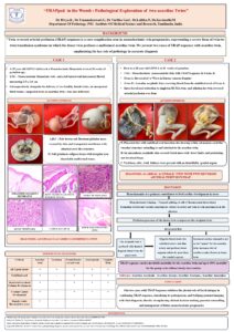
Poster 15: “TRAP”ped in the Womb : Pathological exploration of two acardiac twins
Two cases of TRAP sequence in monochorionic twins highlight the importance of early antenatal Doppler for timely diagnosis and prognosis. Fetal autopsy confirmed absent cardiac structures and severe malformations, reinforcing its vital role in accurate diagnosis.

Poster 16: Uterine dedifferentiated mesonephric like adenocarcinoma- A rare case report
Mesonephric-like adenocarcinomas (MLAs) are recently recognised in the World Health Organization 2020 classification and are regarded as an aggressive subtype of endometrial carcinoma. We report an unusual case of uterine MLA in a 56-year-old woman who underwent hysterectomy and bilateral salpingo-oophorectomy , lymph node dissection and omentectomy for a dedifferentiated MLA.
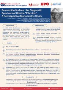
Poster 17: Beyond the Surface: Clinicopathological complexity and diagnoses spectrum of uterine “fibroids”. A retrospective monocentric study.
The 2020 WHO classification expanded the spectrum of uterine mesenchymal tumours beyond the traditional benign–malignant distinction. Among 300 cases, IHC (67.6%) identified 10% non-typical entities. Increased awareness may improve recognition, risk stratification, and surgical management.
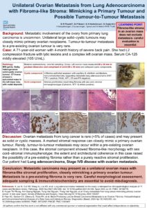
Poster 18: Unilateral ovarian metastasis from lung adenocarcinoma with fibroma-like stromal proliferation: A diagnostic mimic of primary ovarian tumour
This case highlights a potential diagnostic pitfall, as metastatic carcinoma may present as a unilateral ovarian mass with exuberant stromal response, closely mimicking a primary ovarian tumour. Careful morphological assessment, adequate sampling, and appropriate immunohistochemistry are essential to distinguish metastatic disease from primary ovarian neoplasia and to avoid misdiagnosis.
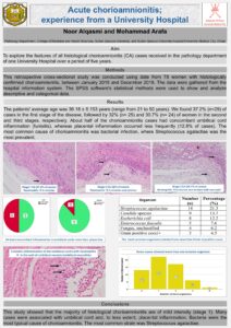
Poster 19: Acute chorioamnionitis; experience from a University Hospital
The majority of chorioamnionitis are mild (stage 1). The most common associated bacterial strain is Streptococcus agalactiae.

Poster 20: Endometrial polyps; experience from a University Hospital
Most endometrial polyps are benign and single with a good concordance between the clinical suspicion and the histopathological confirmation.
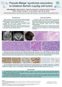
Poster 21: Pseudo-Meigs’ syndrome secondary to bilateral Sertoli–Leydig cell tumor
Meigs’ syndrome is a rare clinical condition defined by the triad of a benign gynecological tumor, such as fibroma or thecoma, ascites, and pleural effusion, resolving after tumor removal. Pseudo-Meigs’ syndrome presents the same features, but is associated with other tumors, including malignant ovarian neoplasms, teratomas and uterine leiomyomas. Sertoli-Leydig cell tumors are uncommon, with only three cases described in the literature in association with Pseudo-Meigs’ syndrome. Case of a 54-year-old woman with bilateral pleural effusion, prominent ascites, bilateral large solid–cystic ovarian lesions and multiple lymphadenopathies.

Poster 22: Undifferentiated uterine sarcoma with HER2 amplification and S100/SOX10-positivity – a new entity?
Uterine sarcomas comprise a heterogeneous group of rare malignant mesenchymal tumors, with high-grade and undifferentiated categories posing significant diagnostic challenges. Case of a 56-year-old woman with abdominal pressure, pain, and urinary frequency. Imaging revealed a markedly enlarged uterus with multiple nodular masses and extensive areas of diffusion restriction, compatible with sarcomatous transformation. Additional findings included multiple lung, liver, pancreas and bone metastases. Biopsy of the left uterine horn was performed.
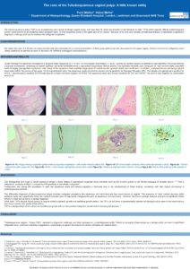
Poster 23: The case of the tubulosquamous vaginal polyp: A little known entity
Rare & deceptive! Our poster explores the tubulosquamous vaginal polyp. This diagnostically challenging biphasic benign postmenopausal lesion can mimic squamous cell carcinoma.
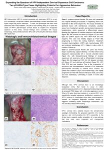
Poster 24: Expanding the spectrum of HPV-independent cervical squamous cell carcinoma: Two p53-wild-type cases highlighting potential for aggressive behaviour
We report 2 HPV-independent, p53-wild-type cervical squamous cell carcinomas, expanding this rare entity beyond the p53-abnormal paradigm and highlighting that non-block p16 plus negative HPV testing may identify clinically aggressive tumors with major management implications.

Poster 25: Squamous cell carcinoma of the cervix associated with low-risk HPV: correlation with an emerging pathogenetic subset
Two cervical squamous cell carcinomas showed isolated low-risk HPV (HPV6 and HPV6/11) with non-block-type p16, supporting an emerging low-risk HPV-associated pathway of cervical carcinogenesis beyond the usual HPV-driven versus HPV-independent dichotomy.

Poster 26: Primary intestinal-type adenocarcinoma of the vagina: clinicoradiologic-pathologic correlation and a stepwise approach to avoid metastatic overinterpretation
Primary intestinal-type adenocarcinoma of the vagina is a rare mimic of metastatic colorectal adenocarcinoma. We report a case with clinicoradiologic-pathologic correlation and propose a stepwise diagnostic approach to avoid metastatic overinterpretation.

Poster 27: Angiogenic and anti-angiogenic marker expression in hypertensive placentas: correlation with placental morphology, doppler findings, and foetal outcome
Placental immunohistochemistry adds value beyond routine microscopy by highlighting VEGF and sFlt-1 expression, helping identify angiogenic imbalance, explain placental vascular lesions, and strengthen correlation with adverse foetal outcomes in hypertensive pregnancies.

Poster 28: A five-year analysis of enhanced pathological examination of sentinel lymph nodes with keratin immunohistochemistry and level sections (ultrastaging) in endometrial cancer at a tertiary referral centre
My poster presents an audit I performed on the diagnostic yield of sentinal lymph node ultrastaging in endometrial cancer and histopathological characteristics predictive of positivity.
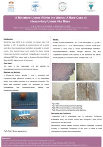
Poster 29: A miniature uterus within the uterus: A rare case of intracavitary uterus-like mass
An unusual intracavitary uterus-like mass highlighting the diagnostic challenge and the essential role of histopathology and immunohistochemistry in differential diagnosis.
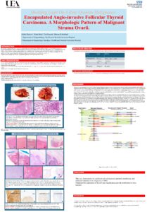
Poster 30: Shedding light on a rare ovarian malignancy: A case of encapsulated angio-invasive follicular thyroid carcinoma
This case highlights the significant role of integrated approach in the diagnosis of MSO as recent studies have shown that they harbour molecular alterations analogous to their thyroid gland counterparts, supporting the application of thyroid-type classification and risk stratification to these tumours.

Poster 31: A rare metastatic pathway from ovary to neck – ovarian sex cord-stromal tumor presenting with Virchow’s node
A rare presentation of an ovarian sex cord–stromal tumour with supraclavicular nodal metastasis.

Poster 32: Differential diagnosis of SMARCA4-deficient tumours in the cervix: A diagnostic journey toward SMARCA4-Deficient Uterine Sarcoma
This case demonstrates the diagnostic complexity of SMARCA4-deficient tumours in the cervix, where distinguishing the exceptionally rare possibility of cervical SCCOHT from the newly recognised SDUS requires close integration of site, morphology, immunophenotype and molecular findings.
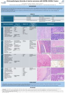
Poster 33: Clinicopathological diversity of uterine sarcomas with KAT6B::KANSL1 fusion
KAT6B::KANSL1 fusion–positive uterine sarcomas represent a newly emerging entity with striking morphological diversity, spanning low to high grade phenotypes and variable IHC expression.
Recognition and genomic testing are essential for accurate classification and management.
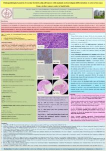
Poster 34: Clinicopathological analysis of ovarian Sertoli-Leydig cell tumors with emphasis on heterologous differentiation: A series of ten cases from a tertiary cancer centre in South India
Rare ovarian SLCTs show marked heterogeneity: 40% heterologous differentiation and 50% higher-grade tumors. Despite early-stage presentation, 20% recurrence highlights need for careful grading, sampling & follow-up.

Poster 35: When limited tissue obscures diagnosis: The role of clinical and morphological correlation in cervical embryonal rhabdomyosarcoma
Embryonal rhabdomyosarcoma (ERMS) of the cervix is a rare malignancy in young women. Diagnosis can be challenging in small biopsy samples. We present a case highlighting the diagnostic value of correlating examination and microscopic findings in limited samples.

Poster 36: Recurrent intestinal-type adenocarcinoma of the vagina with colorectal immunophenotype: primary tumour or metastasis from the colorectum?
A diagnostic pitfall: vaginal adenocarcinoma with intestinal differentiation mimicking colorectal metastasis. Negative systemic work-up and local recurrence over years supports primary origin. Emphasises careful interpretation of immunohistochemistry and clinical context.

Poster 37: Genomic signatures of mucinous ovarian tumours with mural nodules: distinguishing features from mucinous tumours without mural nodules and dedifferentiated mucinous carcinoma
Targeted sequencing of 38 mucinous ovarian tumours shows shared early drivers, grade‑associated genomic complexity, and compartment‑specific alterations in MTMN. DDMC displays a distinct chromatin‑remodelling and BRCA2‑related profile, supporting its separation from MTMN.
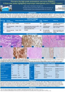
Poster 38: Pilomatrix-like high-grade endometrioid carcinoma (PiMHEC): A report of three cases of a highly aggressive tumour with clinicopathological and molecular findings
3 cases of Pilomatrix -like high-grade endometrioid carcinoma, a rare aggressive variant. Highlights morphologic heterogeneity, CTNNB1 mutation and beta catenin expression. Recognition is essential due to diagnostic pitfalls and aggressive clinical behaviour.
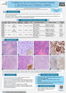
Poster 39: Corded and hyalinized endometrioid carcinoma of the uterine corpus: Report of three cases highlighting a rare morphological variant
3 cases of corded & hyalinized endometrioid carcinoma, including unique combo with pilomatrix-like high grade endometrioid carcinoma & leiomyosarcoma. Recognition vital to avoid pitfalls. Harbours CTNNB1 mutation & nuclear βcatenin+. Can be high stage/aggressive.

Poster 40: Analysing cervical smears using convolution neural network (CNN)
Convolutional Neural Networks (CNNs) demonstrated high accuracy (95%) in categorising cervical smears especially abnormal cytology. However, limited data and overfitting remain challenges. Future work should focus on larger datasets and advanced AI methods such as object detection.

Poster 41: Characterisation of distinct molecular phenotypes in high-grade serous ovarian cancer biopsies to predict neoadjuvant chemotherapy response
Pre-NACT RNA-seq in advanced ovarian cancer identifies distinct transcriptomic profiles linked to Chemotherapy Response Score. CRS3 tumours show differentiated, hormone-related signatures, while CRS1/2 display pro-tumorigenic and immunomodulatory features associated with resistance.

Poster 42: Clear cell carcinoma of the uterine cervix: Report of two cases including a patient with Lynch Syndrome
Two cases of rare, HPV-independent cervical clear cell carcinoma—one in a patient with Lynch syndrome—show variable MMR status. Highlights diagnostic challenges and supports considering MMR testing in HPV-independent tumours where resources allow.

Poster 43: Clinicopathological spectrum of vulvar lesions: An 11 year audit of 634 consecutive cases from a tertiary care centre in India
Vulvar pathology encompasses a heterogenous group of lesions ranging from inflammatory dermatoses to invasive malignancies. Data on clinicopathological spectrum of vulvar diseases from large institutional series in India remains limited. In our study we did an extensive audit of vulvar lesions, the study incorporating 634 patients and study period ranging from 2014 to 2025 (11 years).

Poster 44: Digital pathology as a platform for delivering gynaecological pathology teaching: an evaluation of residents’ perspectives on logistics and ergonomic impact
Evaluating residents’ views on how gynaecological pathology teaching is delivered: Sessions using digital pathology outperformed those with multihead microscopy in ergonomics, flexibility, and ease of use, proving to be an effective and engaging complementary tool.

Poster 45: Haemangiomas of the uterine cervix presenting with abnormal bleeding and cervical mass: Report of three cases including a rare case of epithelioid haemangioma
Cervical haemangiomas presenting with bleeding and mass lesions, mimicking malignancy, including a rare epithelioid subtype. Accurate histology and IHC are key to avoid overtreatment.
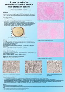
Poster 46: Endometrial stromal tumour with ‘starburst pattern’
Low grade endometrial stromal sarcoma with smooth muscle differentiation showing starburst pattern. 105 mm mass in uterus. Spindle cell nodules with radiating collagenous bands. <1mm from serosa. Positive for CD10, SMA. Negative Cyclin D1, Melan A.
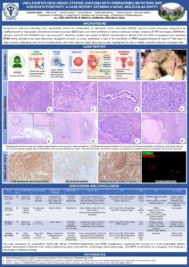
Poster 47: Unclassified high-grade uterine sarcoma with ERBB2/ERBB3 mutations and S100/SOX10 positivity: First report from India of an emerging molecular entity
First report from India of an emerging uterine sarcoma subtype with diffuse S100/SOX10 expression and HER2 amplification. Recognition using morphology, IHC, and HER2 FISH is key due to potential benefit from HER2-targeted therapy.
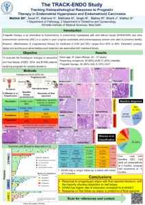
Poster 48: The TRACK-ENDO (Tracking Response to Anti-progestin/Conservative therapy in Kindred ENDOmetrial lesions) Study: tracking histopathological response to progestin therapy in endometrial hyperplasia and endometrioid carcinoma
Progestin therapy offers a fertility-sparing alternative in EHA/EHWA & EEC, with 60–80% effectiveness. Most cases resolve on follow-up biopsy. EHWA shows better outcomes than EHA/EEC, and early response results in favourable outcome.
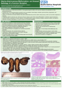
Poster 49: Uterine arteriovenous malformation—an unusual aetiology of a common symptom
Uterine AVM is a rare, potentially life-threatening cause of heavy bleeding that can mimic endometriosis. Histology showed thick and thin walled vessels in myometrium/cervix. This case emphasises the need for accurate preoperative diagnosis to prevent severe haemorrhage.

Poster 50: Metastatic angiosarcoma arising in mature cystic teratoma of the ovary: A rare case report on rare malignant transformation
Beyond the usual suspects: Angiosarcoma arising in mature cystic teratoma.

Poster 51: The surgical diagnosis and distribution of superficial endometriosis: a retrospective analysis of 516 cases, the “40:30:20:10” rule, and use of CD10 immunohistochemistry in histological diagnosis
A retrospective analysis of 516 cases demonstrates that combining laparoscopic excision with CD10 immunohistochemistry enhances diagnostic yield and characterises the multisite distribution of superficial endometriosis (“40:30:20:10 rule”).
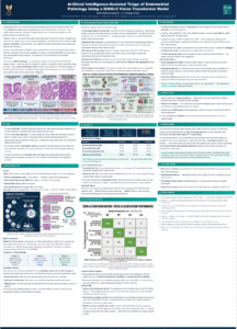
Poster 52: Artificial intelligence -assisted triage of endometrial pathology using a DINOv2 vision transformer model
This study investigated whether artificial intelligence (AI) could assist in triaging and classifying digital endometrial pathology slides.

Poster 53: Weakly supervised AI for ovarian cancer subtyping and tumour localisation in whole slide histopathology
Accurate ovarian cancer subtype classification and tumour localisation can be achieved by weakly supervised AI models trained using whole-slide-level labels alone. This offers explainable-AI decisions which highlight regions of interest in the slide and enables realistic AI deployment in clinical pathology.
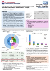
Poster 54: A retrospective audit of the distribution and clinicopathological association of endometrial cancer molecular subtypes
Retrospective audit of the molecular subtypes of endometrial carcinoma in a tertiary referral centre and correlation with histological subtype and FIGO stage.